Poznámky k bionomii Farsus dubius (Piller a Mitterpacher, 1783) (Coleoptera: Eucnemidae: Melasinae), s pozorováními jeho hypermetamorfózního vývoje
Tamás Németh & Robert L. Otto
1)Hungarian Natural History Museum,Coleoptera Collection, H-1088 Budapest, Baross utca13, Hungary
E-mail: haesito@gmail.com
2)2301 Cypress Way, Apt. 22, Madison, WI 53713, U.S.A.
E-mail: tar1672@yahoo.com
Abstract – Additional information to the biology of Farsus dubius (Piller and Mitterpacher, 1783) is presented. Perris’s descriptions and observations are compared, different developmental stages are photographically imaged. With 22 figures.
Key words – False click-beetles, saproxylic, Palaearctic, hypermetamorphosis, fifth instar, prepupal larval forms
INTRODUCTION
Farsus Jacquelin du Val, 1863 is a small, primarily Old World group, consisting of 12 described species, eight of which are distributed in the tropics and the remaining four are in the Palaearctic Region. Farsus brevis Fleutiaux, 1931, Farsus exoticus Bonvouloir, 1875 and Farsus salvazai Fleutiaux, 1918 are distributed in the Indo-Malayan ecozone. Farsus costipennis Fleutiaux, 1926 have been taken in Madagascar. Farsus ainu Fleutiaux, 1922 is distributed in Japan. Farsus obenbergeri Roubal, 1941 have been taken in Sichuan, China. Farsus obesus Iablokoff-Khnzorian, 1964 is regarded as a precinctive species in Georgia. The type species, F. dubius (Piller and Mitterpacher, 1783) have been recorded in many countries across western and central Europe. Muona (1991) noted two undescribed species are present in S.E. Asia. Muona (2000) also noted the clade consisting of four species (Farsus elavatus Bonvouloir, 1875, Farsus oblitus Horn, 1890, Farsus obscurus Bonvouloir, 1875 and Farsus wagneri Fleutiaux, 1934) from Central and South America are misplaced. These four species currently remain in Farsus, until a new group name is proposed by Muona. Muona (2000) removed one species, Farsus convexus Fisher, 1945 and placed it in his new genus, Protofarsus, and described Protofarsus caribicus from the Nearctic Region.
Farsus, Protofarsus, Arrhipis Bonvouloir, 1875 and Sarfus Fleutiaux, 1925 each have produced elytral apices. Farsus is distinguished from Protofarsus by its caudally obliterated lateral elytral ridge. Lateral elytral ridge is complete in Protofarsus. Farsus is further distinguished from Arrhipis and Sarfus by the apices of the frontoclypeal region in relation to the base. Apices of the frontoclypeal region are more than twice as wide in Farsus, and less than twice as wide in Arrhipis and Sarfus.
Current distribution of Farsus dubius – Albania (Mertlik 2011); Czech Republic, France, Hungary, Spain, “Yugoslavia” (Muona 2007); Austria, Italy, Romania, Bosnia and Herzegovina, Caucasus, Greece, Montenegro, Syria, Turkey (Horion 1953), Bulgaria (Mertlik et al 2009).
Horion (1953) published the first records of F. dubius from Syria, Bosnia (Urac) and Herzegovina (Igbar) as well as Turkey. Burakowski (1991) erroneously mentioned the first record of F. dubius from Syria. Mertlik and Platia (2008) also erroneously listed F. dubius as a new record in Turkey. Additionally, Mertlik et al. (2009) also published the first record for the eucnemid species in Bosnia and Herzogovina in error. Horion (1953) published the first and only data from Montenegro: “Montenegro (Mustajbeg)”.
MATERIAL AND METHODS
Specimens examined – Hungary: Baranya megye [county], Kétújfalu, lámpán [at light], 26.VII.1987, leg. József Sár (1 specimen); Veszprém megye [county], Zalahaláp-Ódörögd, Csilla-hegy, tölgyes [oak forest], egyelés [hand-collected], 18.VI.1993, leg. Péter Bartha (2 specimens); Veszprém megye [county], Zalahaláp, Csilla-hegy, from rotten Quercus, 7.VII.2013, leg. A. Kotán & T. Németh (16 specimens); same locality, but 17.VIII.2013, leg. E. Jókuthy, O. Merkl, P. Nemes & T. Németh (36 specimens); same data, but reared from rotten Quercus 17.VIII.2013–20.V.2014 (40 specimens). Montenegro: Bar distr., Mt. Vrsouta, Sutorman, mixed forest, from rotten Fagus, 42°9'18.32"N, 19°5'52.81"E, adult cadavers, 1–2.VI.2014., leg. V. Dušánek, R. Kundrata & T. Németh; same data, but reared from rotten Fagus, 2.VI.2014–10.VII.2014 (2 specimens).
Rearing – Pieces of wood containing larvae and pupae were placed in plastic boxes during the winter season. Boxes were left open for a short time to dry out the pieces to assure mould would not grow on the surfaces. These boxes were maintained at a temperature of 23 °C during the entire time.
Preparation of Larvae and Adults – Larvae and pupae were directly immersed into vials filled with 70% ethanol. Alcohol was changed once after initial immersion. Adults were mounted on paper cards. Larvae and adults are deposited in the Coleoptera Collection of the Hungarian Natural History Museum, Budapest and the collection of the Global Eucnemid Research Project (GERP) at the Department of Entomology, University of Wisconsin – Madison, U.S.A.
Translations – The entire section by Perris (1871) in Bonvouloir’s monograph was retranscribed in a Word document. The section was copied and pasted onto a translation website, http://www.bing.com/translator and later verified through another translator site, http://www.translate.google.com. The translated section was copied and pasted back into the word document to be analysed later. Most of Perris’s account on his observations (i.e. analysis on nutritional sources, debating on segmentation numbers, etc.) were omitted to focus mainly on his descriptions of both larval forms and pupae. Adding relevant section of Perris’s work is important, in order to have all known bionomic information of F. dubius, by comparing similarities and differences of his and our observations. This text would also be interesting in the point of view of the 19th century entomology.
QUOTATIONS FROM PERRIS’S (1871) ACCOUNT
“Sometime around May 1842, I found several dead bodies of the scarce insects, then called Farsus unicolor beneath the thick bark of an old dead oak tree near Mont-de Marsan. They probably came from the tree that I was exploring; but I looked in vain for signs of larvae or a living individual, and made even since that time, unnecessary research on dead oaks that fell into my hands.
More than twenty years later, in 1862, M. Bauduer, a young entomologist full of ardour, living in the town of Sos (Lot-et-Garonne) has sent me an almost rotten fragment of an oak trunk, riddled with holes and galleries what I initially believed to be the work of Platypus cylindrus, but which, observed with some care, allowed me to see a large number of Farsus cadavers. Without worry of the event which the generation perished before emerging, I came to a certain point of view, which would allow me to more positively solve the habitat question of larvae of this insect.
The following year, Mr. Bauduer was happy in which he collected more than 300 Farsus when he had the chance to find a long oak trunk lying on the ground, partly reduced to an almost spongy state; so it is true that perhaps the insect is not rare , and when its way of life or its breeding site is discovered, it can be obtained in quantity, as I experienced it many times. Mr. Bauduer quickly sent me large very productive tree sections, and, in exploring, I found hatched Farsus pupae and some larvae which form corresponds well to the idea that I made and other very much different larval forms that strongly piqued my curiosity. At first sight, I took them to be parasitic wasp larvae.
Their very smooth surface, the smallness of their head, the simplicity and conformation of the mouth organs seemed to justify this opinion; but it was contradicted by the number of segments and the position of the stigmata. I was therefore very intrigued. I reported these larvae to M. Bauduer, with an invitation to make every effort to follow the metamorphosis, and I promised to do so on my side. Not having the time to study them, I put several in alcohol, as I had already done for recognised nymphs and larvae belonging to Farsus, in order to review it later.
This happened in July 1863. Since that time, no Farsus was born from my wood until July of the following year. In desperation, I started to butcher these woods and I found more of these larvae which I was so concerned about. Mr. Bauduer had not learned more than I. On my request, he sent me a new stretch of oak; I found two kinds of known observed larvae, living in cells or separate galleries, without that one seemed to be parasitic on the other. By studying them so carefully, I found them, despite differences of sliced form and large differences of structure on the side of the head, approximation points that persuaded me that the larva, seemingly Hymenoptera, was only a first state of the larva of the Farsus. To control this assumption, I assiduously followed their development, discovering from time to time some of them, and I finished by noting that larvae object of so many doubts and concerns took the form of those which I was perfectly safe.
Now I give the description of these two larvae.
PRIMARY LARVAL FORM OF FARSUS
Maximum length: 7 millimeters. Elliptical, very bulbous, legless, white almost matte, fleshy, pretty firm, perfectly hairless and smooth, even under the microscope.
Head very small, embedded in the prothorax, slightly longer than wide, subconvex, white like the rest of the body, marked by three red lines coming together slightly above the middle; the two lower ones arched, meeting together forming a half an ellipse, the upper bifurcated towards the vertex. These lines are a little thicker at their meeting point, where the lower seem very strong under a magnifying glass, forming a kind of sharp blade, partially enclosing a fleshy nipple that seems overcame sub-horny tubercles. It is in this state of simplicity that arise, even at high magnification, the structures that make up the mouth; but under the microscope we see something else. This instrument shown in fact is the aforesaid nipple that supports a conical end to a bifurcate tongue, and right and left of this strip is a small appendage which is slightly shorter, more hail, simulating a uniarticulate palp and having probable functions.
The body, very bulbous, as I said, but a little depressed, is composed of twelve segments, including three for the thorax and nine for the abdomen. All these segments are roughly equal in length; but it goes without saying, given the form of the larva, it is far from being the same for the width. The last segment is regularly rounded, without the slightest crease. The sides are not retractable, but they are scalloped by the protrusion of the segments. On the middle of each segment, both above and below, in part however the prothorax and the last abdominal segment, one perceives any small plate or russet areola as sub-horny, at least on its perimeter ellipse in cross-section, are not retractable and are stigmatiform. One is, indeed, tempted to take these areoles for stigma, despite the anomaly of their number (10 pairs) and their position in all segments except the first and the last, if it was struck by the sight of the true stigmata, very apparent along the flanks. They are russet, circular, well marked peritreme, and number nine anterior edge of mesothorax and one near the front edge of the first eight abdominal segments.
With the regularity of shapes and details, and the small appearance of the head structures, it would be difficult to tell at the first glance, where are the top and bottom of this larva, since these two faces look perfectly. If considered aside, we note that, on the one hand, it is quite strongly convex, and the other very-little convex and somewhat sinuous. Naturally one would believe that the convex side is back, and it would be a mistake, because it is precisely that of belly. Or recognized the anus located on this face, very-near the base of the last segment. It has the depressed, almost semi-discoidal form, with a median fold.
SECOND FORM OF LARVA
If you look at the figure that I gave of the second larva and compare it to that of the first, we cannot help but think that they belong to two very different insects; and we will dwell on this comparison, more we will confirm this conviction, because the details are no less diverse as the whole. They have these in common: 1st the absence of legs; 2nd the number segments, which is twelve, insignificant character for beetle larvae, but that led me to this conclusion that the bulbous larva was not the class of parasitic Hymenoptera larvae, which are known to me, have thirteen segments; 3rd the number, position and shape of the stigmata; 4th small, elliptical areoles, both above and below, in the middle of all segments, except the two extremes. This last feature, the Farsus larva offered me the first example, and must be very rare, appeared to be very important to me, in which became the determining ground of reconciliation that more observation was later confirmed.
Now let's see what changes has undergone from the first larva to become the second.
Its maximum size of 7 millimeters could reach the 10-11 millimeters. Its form, elliptico-bulbous and depressed on the back side, becoming elongated, almost cylindrical, with a sensitive and rather abrupt narrowing previously, gradually decrease in diameter towards the posterior end. The body remained smooth, but its once perfectly smooth surface even at the microscope, is covered with very small, very tight, tilted back, spinuliform asperities. It occurred, both above and below the abdominal segments, uneven, wavy lines pointing obliquely towards the base or sides. Its fleshy head convex, very small and inconspicuous, marked with reddish lines as mentioned before, with the mouth organs barely visible and not protruding except the palps, which has changed into a sort of like a prism or snout, ferruginous, firmly horny; all of a piece, convex on one side with a few inconspicuous inequalities, concave on the other with a fine edge in the middle, some longitudinal grooves shortened, and two base elevations, two triangular apexes that increase the concavity. The anterior edge is deeply cut with six teeth, separated into two groups of three, with the median, which is longer, wider and slightly obliquely truncated, and the other two triangular. The red lines on the head have completely disappeared, and are transported on the first segment, where they form, both above and below, an inverted V with slightly curved and slightly downward branches. Between these two branches, the side where the head is concave, arises a a pretty big fleshy nipple, slightly retractile and obsoletely lobed.
I said that I've proven the difficulties to recognize the back and belly of the first larva. I thought I could speak without hesitation for the second. Obviously, I have said, the ventral region is on the side where the head is concave. The larva, indeed, feeding on wood, it is natural to think it is acting from top down, drawing in woody tissue using the instrument that nature gave him, and that therefore the underside of the head is concave face; because it is on this side only that the instrument seems to be able to operate with some energy as a pick or rake. I nevertheless wanted to check the thing, and this is not without a big surprise that I met the anus on the side where the head is convex. but the anus was exceptionally on the side of the back? Nothing was impossible with a larva that had other excepts to the ordinary rules. I then started to explore the head with the greatest care and with the help of a very strong magnifying glass. On the concave side, I've found that what I have already said, and therefore nothing that could shed light on the question. On the convex side, on the contrary, I noticed distinctly, in the deep indentation left by the two groups of teeth, a small piece of rectangle area, followed by an another smaller area and previously subrounded. Well, have I exclaimed, an epistome and a labrum; so that’s the top: it is the bottom, told me at once three small appendages surmounting the second area, which became, therefore, in my opinion, analogous to that which I had already observed in the first larva. I had in fact, see in these bodies either a epistome and labrum, but a chin, lip, tongue and palps that I cannot simply consider as places on the front.
The morality of these hesitations, these assumptions and these final solutions is that it is nescessary to have a fairly large number of individuals to see in one what the other hide and control what we saw or thought I saw for the first time, and that there are still disadvantages to studying dead subjects whose bodies are not moving, and preserved in alcohol, which these organs contracts and often prevents one from properly seeing the composition and structure.
My investigations have revealed to me something else. On the same convex face of the head, an oblong cavity exists below the angles between each side, external from a conical tooth of the broad sill tooth. This cavity shown under very-high magnification, is tuberculiform, a membro-fleshy body, composed of two retractable sections, of which the second is surmounted by two small points. What are these bodies? If they were much closer palps already reported and labial palps as are set on a lip, I would describe as them maxillary palps; but obviously they are not part of the mouth accessories, and therefore I call them antennae. It is true they are not inserted as usual to the lateral angles of the head; but it is that this anomaly has something very surprising in a larva which offer so many others?
Galleries are cylindrical as its body, and therefore no special characters, and it likes soft, spongy wood, probably because the oak that it nourishes is much harder than the light? Here again we see galleries perpendicular to the wood fibers more or less filled with powdery droppings, and completely lacking remains of larvae of other species. Oak fragments locked in a bag gave me many Farsus, and nothing more.
We know that the Farsus larvae are legless, and we have seen that they have no pseudopods, nor exhibit these beads in both wood-destroying larvae. We must admit, there are no organs of locomotion to very energetically browse a gallery of 4 centimeters in length in two years; but it is nevertheless necessary in order to fight against the resistance of the wood apart and they have to grind, they can grow stronger and have strong support points. Nature has not denied the means, and I see clearly evident in these small plates or lipped edge to sub-stratum that carry the segments above and below, in the form of expandable and bulbous body of the primary larval form; and for those of the second form, in the fleshy extractile tubercule located on the dorsal surface of the first segment, especially their body is covered in the countless tilted back asperities. When they move, their wrinkled body expands, it clings from all sides to smooth walls of the gallery, and the prothoracic tubercule probably serves as a special fulcrum working to rake and relief for action of this lever. How easy and how effective combinations! What solicitude for the larva, apparently so deprived, which works in the shadows for nearly two years and should never see the day!
It is in the same galleries where Farsus larvae transform into pupae.
PUPAE
It is naked, that is not shrowded in a shell, and it presents all the parts that form the perfect insect. Figure I give, provides me any description I will say only that there is some extremely fine hairs on the prothorax, along the flanks and the rear edge of the last segments of the abdomen, and varies in size from 3 to 10 mm.
The insect exited through a perfectly round hole.”
OBSERVATIONS
Hungary – Most species of false click beetles are rare in Hungarian collections. Faunistic research focusing on saproxylic beetles resulted in more information about these uncommon species. In Hungary, Farsus dubius was previously known from four areas. One specimen is known from Baranya county in southern Hungary (Sár 1992); two data were mentioned by Kuthy (1897) from the Mátra Mountains in Parád and from Vajszló in southern Hungary. Two specimens were collected more than two decades ago in Veszprém county, western Hungary. With the kind help of Péter Bartha, who collected these two specimens during his military service on 18 June 1993, we found an abandoned military training area (Fig. 1). The site was first visited on 10 July 2013, then on 17 and 23 July with amateur collectors. The habitat is a wooded pasture with open oak groves and solitary trees about 50–100 years (Q. cerris, Q. petraea, Q. pubescens), some of which are dying or have fallen. Each visit was successful in obtaining F. dubius specimens. After a short daytime search, we found dead oaks 30 centimeters in diameter. Aggregation of beetles was observed hiding beneath the loose bark of dead sessile oaks (Quercus petraea (Mattuschka) Lieblein). After sunset, these beetles were rapidly running on the fungusy parts of the log and started mating. Some females were hiding in the crevices of the log and began laying eggs. We cut a section of the log using a saw. Sections of the log were kept in large plastic boxes.
While opening these pieces during the winter season variously sized larvae (Fig. 2) were present in the softest zone of the wood, 1 cm deep from the surface. Softer parts were occurred around crevices, where water could leak inside the log. Strange, maggot-like “sacks” were observed among small and larger larvae in the soft wood, which looked like desiccated inactive larval cadavers, but were alive and slowly moving.
The first adult began to emerge from the log on 11 May 2014. Intensive swarming took place in the subsequent week with dozens of additional beetles emerging from the collected section of the log. The last adult emerged on 1 June 2014 (Fig. 3). Some specimens were observed mating (Fig. 4) and females began to lay eggs. Round emergence holes appear in clusters and varied considerably in size from 1.5–3.0 mm in diameter.
After carefully opening the wood pieces again in Mid-August of 2014, eight additional larvae were found in the completely dried out sapwood. Just as observed during the winter months, sack-like larvae were present inside their pupal chambers. Outer parts of the exuvia adhered to the walls of the pupal chamber. These larvae appeared to be cadavers. Parts of their exuvia sloughed off while removing some larvae from their chambers (Fig. 5). Remains of white fungus were present on the cuticle of the larvae (Fig. 6). The extracted larvae slowly moved following their removal from the pupal chambers.
Montenegro – During a collecting trip in the Balkan Peninsula in 2014, several adult F. dubius cadavers were discovered beneath the bark of ancient large beech log (Fagus sylvatica L.) in the Rumija Mountains, Montenegro (Fig. 7). The habitat was different from the Hungarian locality, with shady and wet mixed beech forest. With the discovery of numerous last season’s adult cadavers in the old beech log, confidence remained positive to locate immature stages in the sapwood. After carefully removing the bark from the wet, fungusy log, pupae and larvae were present in the soft wood, just beneath the thin bark (Fig 8). Additionally, many strange “sacks” were also present in the wood. These larvae resembled parasitoid wasp larvae rather than beetle larvae (Figs 9 and 10), just as Perris had observed before. Sections of the log containing pupae (Fig. 11) and “sacks” were collected and placed in a box. Most larvae molted into pupae during the remaining leg of the Montenegro trip. These “sacks” or 5th instar larval forms (Fig. 12 and 13) observed to change as well, molted into fully grown prepupal larval forms (Fig. 14 and 15) during the collecting trip. Following the trip to Montenegro, some specimens were placed into small paper tubes, and subsequently into a plastic box with collected parts of the beech log. After four weeks, fully grown prepupal larval forms molted into pupae. Adults began to emerge from the paper tubes and beech log after the following week.
DISCUSSION
Head capsule – It is clear from the descriptions provided by Perris that he described the first form of the larva (Figs 16 and 17) as the 5th instar larval form. The second form he later described (Fig. 18) is the prepupal larval form. The senior author extracted a larval cadaver (Fig. 19) from a pupal chamber, midway through its molting process. The illustration clearly shows a transition between the 5th instar and prepupal larval stage in the lifecyle. The exuvial remains of the former head capsule can be seen on the dorsum as the prepupal larval form attempted to squeeze out of its exuvia. That observation clearly shows the sequence of development in the species’ lifecycle.
The head capsule in the 5th instar stage (Fig. 20) is much more different, when compared with the prepupal larval form (Fig. 21). It is more simplified, fleshy with a pair of sclerotised lines on one side and a sclerotised forked line on the opposite side. Based on examining a number of preserved specimens, it appears the head is eversible, often hidden within the prothorax. Although it possesses ventral mouth parts in the buccal region, it is unknown how the larval form obtains nutrition in the pupal chamber, possibly through extraoral digestion outside the larvae’s body as described by Muona and Teräväinen (2008). The head capsule of the prepupal larval form is much more prominent and complex, well armed with six anteriorly directed lateral projections (Fig. 21).
Exuvia – The presence of these extremely delicate, paper-thin exuviae wrapped around the 5th instar larvae and prepupal larval forms in the pupal chamber is a new observation never recorded by Perris as he studied and described larvae of F. dubius over 140 years ago. This phenomenon was reported one other time as Gardner (1935) wrote, “with the above are four larvae of peculiar structure; the removal of the skin of the largest of these exposed a larva of the later form as already described” as the collector obtained mature larvae of Arrhipis orientalis Fleutiaux, 1896 in the sapwood in India. Observations made by the collector while obtaining larvae of A. orientalis in India is very similar to the senior author’s observations of collecting F. dubius in Hungary and Montenegro. No other observations have been recorded elsewhere in the family, but it does not mean the phenomenon is non-existent in other groups. It is quite likely the phenomenon may be present in other groups within the family. Many encountered larvae, especially in the tribe Dirhagini were found in pupal chamber devoid of the exuvia that would otherwise line against the wall. Nearly all larval types were found doubled up in a U-shaped fashion inside the chamber. With its fusiform design, F. dubius is incapable to double up inside the pupal chamber as observed in majority of other encountered eucnemid larvae.
The purpose of exuvia lined againts the walls of pupal chamber is unknown. It may act as a protectant when outside conditions are at its most unfavourable, like dry autumn, or wet winter in order to protect the vulnerable stage before springtime pupation. It may also protect the pupal chamber from dessication and/or excessive moisture that would lead to undesirable fungal and bacterial growth. Another possibility is, it may not serve a purpose at all. Further research is needed to understand this unique situation present in Farsus.
Fusiform larvae – Most commonly within Eucnemidae, either elateriform or buprestiform larvae are often encountered (see Peterson 1951, Leiler 1976, Mamaev 1976, Otto 2012a, 2012b, 2013, 2014, 2015, Otto & Gruber 2016, Otto & Young 1998). Farsus are among a few groups within the family, whose larvae have a fusiform body design. Fusiform body design is present in known larvae of Arrhipis, Galbimorpha and Galbites. Gardner (1935) described and illustrated larvae of both Arrhipis species and Galbimorpha agasterceroides (Fleutiaux, 1896). Suzuki (2012) described larvae of Galbites borealis (Hisamatsu, 1980) collected in Japan. All fusiform larvae are less sclerotised, with much wider than long segments, often resembling dipteran or hymenopteran larvae. Areoles are usually present. Microtrichial patches are not arranged in elaborate shapes on any segments as compared to other larval forms. Microasperites are usually present on surfaces of the thoracic and abdominal segments. The head is disproportionately smaller, simple, often armed with four to six anteriorly directed projections. Mandibles are not present, but a buccal orifice is present on the ventral side of the head.
Onisciform larvae – Gardner (1935) illustrated and described the larva of Arisus bituberculatus Fleutiaux, 1935. It is the only larval type in which the 8th abdominal segment laterally encloses the 9th abdominal segment. That larval type, along with larvae of Dorhn’s Elegant Eucnemid Beetle, Paleoxenus dohrni (Horn, 1878) in the Nearctic region and Old World species of Dromaeolus (i.e. Dromaeolus barnabita (Villa, 1836)) represent a fourth form, onisciform, which is very rarely present in the family. The larval form is much more dorso-ventrally flattened compared with the other three larval forms, along with a usually well sclerotised exoskeleton and wider than long thoracic and abdominal segments.
Hypermetamorphosis – In the Palaearctic region, the number of species with multiple larval forms may be around 20 species, but may be higher as further discoveries are made. Hypermetamorphosis varies greatly throughout the family in different stages of development involving either first instar free-living triungulins as in Dirrhagofarsus attenuatus (Mäklin, 1845) (Burakowski 1989) or prepupal larval forms (i.e. Farsus and Eucnemis) or both (i.e. Nematodes, Otto pers. obs.). Muona (1993) wrote that hypermetamorphosis is common in the tribe Dirhagini, subfamily Eucneminae and to some degree is present in the tribe Nematodini. The junior author found a prepupal larval form for the macraulacine genus Asiocnemis in the San Gabriel Mountains, just north of Los Angeles, California, U.S.A., which may indicate some genera within the tribe Macraulacini may also undergo some degree of hypermetamorphic lifecycle as well. Prepupal larval form of Asiocnemis is somewhat similar to mature larvae in form, but less sclerotised, lacking areoles and microtrichial patches. Head capsule and last abdominal segment are structurally different than the 5th instar larval form. Fifth instars of Asiocnemis have three lateral projections along each side of the head capsule. Prepupal larval forms lack any lateral projections along each side (Otto pers. obs.).
In Isorhipis, Melasis and Nematodes, prepupal larval forms are usually shorter in length compared to the 5th instar larval form. Additionally, areoles, microtrichial patches and prothoracic scleromes are often reduced or absent altogether. The head capsule undergoes very little structural changes as 5th instar larvae transitions into a prepupal larval form as observed in these genera. Burakowski (1989) described the hypermetamorphic lifecycle of D. attenuatus then called Rhacopus attenuatus collected in Poland. He observed several different larval forms for the species as he studied it. These different forms (1st instar triungulins, 2nd instar and 5th instar larvae) each differ by the structures of the mandibles, head capsules and general form of body.
In Eucnemis, prepupal larval stage take on a different shape compared to the 5th instar. Fifth instars of E. capucina generally look like a typical melasine or macraulacine larva with well sclerotised exoskeleton, prothoracic scleromes, microtrichial patches and possess seven lateral teeth or projections on each side of the head capsule. Prepupal larval stages are less sclerotized with reduced prothoracic scleromes and are shorter in length. Microtrichial patches are also reduced or absent altogether. Head capsule takes on a different shape compared to the 5th instars. Head capsules in prepupal larval stages are generally much smaller, more triangular, fewer lateral teeth or projections along each side of the head which are directed anteriorly and are more pronounced near the anterior end.
Fifth instar larval form of F. dubius is very similar to A. orientalis in India, including structures of its simplified head. By comparing information and illustrations 22 and 23 on plate 2 (Fig. 22) in Gardner’s (1935) paper with F. dubius larvae, the only difference that can be observed is the size of the areoles (larger in F. dubius; smaller in A. orientalis) and presence/absence of areoles on the prothoracic and 8th abdominal sternum (present in F. dubius; absent in A. orientalis). Additionally, further comparisons of the prepupal larval form of F. dubius against prepupal larval forms of A. orientalis (illustration numbers 19–21; plate 2) indicate further means to differentiate these species. The structure of the head capsule, along with the shape of the prothoracic scleromes may separate these species.
As mentioned earlier, only a small number of larvae are known for Eucnemidae. It is just a tip of the iceberg and many more new discoveries and new observations await to be uncovered as we continue to explore the natural world. Future new discoveries on life histories and new species will lead to new, additional information that will change how we view this unique coleopterous family and the role it plays in the forest communities around the globe.
Acknowledgements – We thank Jyrki Muona for taking time to answer some questions we had during the course of this project. The senior author would like to thank the opportunity to collect for Václav Dušánek, Attila Kotán and Robin Kundrata. Thanks are due to Ottó Merkl for text review, and valuable advice for the manuscript. Special thanks goes to Péter Bartha and Nikola Rahmé, for providing the exact locality data for the largest known Hungarian population of Farsus dubius. We thank Paul J. Johnson for offering his input on the manuscript.
REFERENCES
BURAKOWSKI B. 1989: Hypermetamorphosis of Rhacopus attenuatus (Maeklin) (Coleoptera, Eucnemidae). – Annales Zoologici 42(5): 165–180.
BURAKOWSKI B. 1991: Klucze do Oznaczania Owadów Polski. Czesc XIX. Chrzaszcze Coleoptera. Zeszyt 35–37. Cerophytidae, Eucnemidae, Throscidae, Lissomidae. – Polskie Towarzystwo Entomologiczne, Warsaw, 91 pp.
FAUNA EUROPAEA 2010: Fauna Europaea version 2.3. – Available online at http://www.faunaeur.org. [Accessed 30 April 2016.]
GARDNER J. C. M. 1935: Immature Stages of Indian Coleoptera (17) (Eucnemidae). – Indian Forest Records 1(4): 79–93.
HORION A. 1953: Faunistik der mitteleuropäischen Käfer. Band III. Malacodermata, Sternoxia (Elateridae-Throscidae). Entomologische Arbeiten aus dem Museum G. Frey, Tutzing b. München, 340 pp. Eigenverlag Museum Frey, München, Entom. Arb., Museum G. Frey München, XIX Sonderband, 340 pp.
KUTHY D. 1897: Ordo. Coleoptera. – In: A Magyar Birodalom Állatvilága (Fauna Regni Hungariae). III. Arthropoda. (Insecta. Coleoptera.) Királyi Magyar Természettudományi Társulat, Budapest, 213 pp.
MAMAEV B. M. 1976: Morphological types of xylophagous beetle larvae (Coleoptera, Eucnemidae) and their evolutionary importance. – In: Mamaev B. M. (ed.): Evolutionary morphology of wood boring larvae.Nauka, Moscow, pp. 136–155. [In Russian.]
MERTLIK J., JENIŠ I. & ZBUZEK B. 2009: New records on the distribution of some species of the family Melasidae (Coleoptera) – II. – Elateridarium3: 1–6.
MERTLIK J. & PLATIA G. 2008: Catalogue of the family Cebrionidae, Elateridae, Lissomidae, Melasidae and Throscidae (Coleoptera) from Turkey. – Elateridarium 2: 1–40.
MERTLIK J. 2011:New data on the distribution of three species of the family Lissomidae and Melasidae (Coleoptera). – Elateridarium 5: 55–58.
LEILER T. E. 1976: Zur kenntnis der Entwicklungsstadien und der Lebenweisenord- und mitteluropäischer Eucnemiden (Col.). – Entomologische Blätter72: 10–50.
MUONA J. 1991: The Eucnemidae of South-east Asia and the West Pacific – A Biogeographical Study. Australian Systematic Botany 4: 165–182.
MUONA J. 1993: Review of the Phylogeny, Classification and Biology of theFamily Eucnemidae (Coleoptera). – Entomologica Scandinavica, Supplement 44: 1–133.
MUONA J. 2000: A Revision of the Nearctic Eucnemidae. – Acta Zoologica Fennica 212: 1–106.
MUONA J. 2007: Family Eucnemidae. p. 81–86. In: Löbl I. & Smetana A. (eds):Catalogue of Palaearctic Coleoptera, Vol. 4. Elateroidea – Derodontoidea – Bostrichoidea – Lymexyloidea – Cleroidea – Cucujoidea. Apollo Books, Stenstrup, 935 p.
MUONA J. & TERÄVÄINEN M. 2008: Notes on the Biology and Morphology of False Click-Beetle Larvae (Coleoptera: Eucnemidae). – The Coleopterists Bulletin 62(4): 475–479.
OTTO R. L. 2012a: Eucnemid Larvae of the Nearctic Region. Part I: Description of the Larva of Rhagomicrus bonvouloiri (Horn, 1886) (Coleoptera: Eucnemidae: Melasinae: Dirrhagini), with Notes on its Biology. – The Coleopterists Bulletin 66(3): 219–223.
OTTO R. L. 2012b: Eucnemid Larvae of the Nearctic Region. Part II: Description of the Mature Larva of Deltometopus amoenicornis (Say, 1836) (Coleoptera: Eucnemidae: Macraulacinae: Macraulacini), with Notes on its Biology. – The Coleopterists Bulletin 66(3): 285–288.
OTTO R. L. 2013:Eucnemid Larvae of the Nearctic Region. Part III: Mature Larval Descriptions for Three Species of Onichodon Newman, 1838 (Coleoptera: Eucnemidae: Macraulacinae: Macraulacini), with Notes on their Biology. – The Coleopterists Bulletin 67(2): 97–106.
OTTO R. L. 2014:Eucnemid Larvae of the Nearctic Region. Part IV: Description of the Mature Larva of Entomophthalmus rufiolus (LeConte, 1866) (Coleoptera: Eucnemidae: Melasinae: Dirhagini), with Notes on its Biology. – The Coleopterists Bulletin 68(2): 331–335.
OTTO R. L. 2015: Eucnemid larvae of the Nearctic region. Part V: Fifth instar larval descriptions for eight species of Microrhagus Dejean, 1833 (Coleoptera: Eucnemidae: Melasinae: Dirhagini), with descriptions of four new species and notes on their biology. – Insecta Mundi 0421: 1–46.
FIGURE CAPTIONS

FIGURES 1–6. 1 – Military training area in West Hungary, with the largest known population of Farsus dubius in the country; 2 – Living F. dubius prepupal larval form; 3 – Freshly emerged adult specimens; 4 – Pair of F. dubius in copula; 5 – 5th instar larvae, with sloughed-off exuvia; 6 – 5th instar larvae with white fungal growth in pupal cell.

FIGURES 7–11. 7– Fagus forest in Rumija Mountains, Montenegro, habitat of F. dubius; 8 – Collected section of rotten Fagus log containing larvae and pupae; 9–10 – 5th instar larvae in rotten Fagus log; 11 – Pupa from rotten Fagus log.

FIGURES 12–18. 12 – 5th instar larva, dorsal view; 13 – 5th instar larva, lateral view; 14 – Prepupal larval form, dorsal view; 15 – Prepupal larval form, lateral view; 16 – Original drawings of Perris, 5th instar larval form, lateral view; 17 – Original drawings of Perris, 5th instar larval form, dorsal view; 18 – Original drawings of Perris, prepupal larval form, dorsal view.
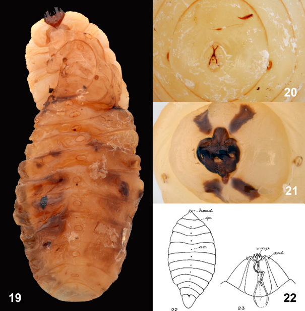
FIGURES 19–22. 19 – Larval cadaver, midway through its molting process; 20 – Head capsule of 5th instar larval form; 21 – Head capsule of prepupal larval form; 22 – Larva of Arrhipis orientalis, original drawings of Gardner, 1935.